Microscopes gradually evolved from eyeglasses and optical lenses that have been in use since about the 13th Century.
 But the modern light microscope was born somewhere in the late 16th and early 17th Century.
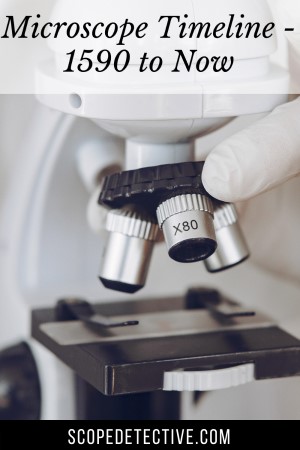
In this post, I’ll outline the evolution of the microscope from the rudimentary lenses of the 1590s through to the sophisticated atom-viewing electron microscopes of today.
Let’s start with a quick run-through of the timeline.
If you’re looking for a timeline of microscope discoveries, see here.
Microscopy Timeline
1590 – Janssen & Lipperhey work on Compound Lenses
1609 – Galileo Develops Microscopes
1665 – Robert Hooke develops Cell Theory
1678 – Van Leeuwenhoek views Bacteria
1729 – Chester Moore Hall invents Achromatic Lens
1846 – Carl Zeiss Begins Manufacturing Microscopes
1870 – Ernst Abbe invents the Abbe Condenser
1893 – August Kohler invents Köhler illumination
1931 – Max Knoll and Ernst Ruska invent the Electron Microscope
1932 – Phase Contrast Microscopy Developed
1981 – Gerd Bennig and Heinrich Rohrer invent the Scanning Tunnel Microscope
1986 – Gerd Bennig invents Atomic Force Microscope
History of the Microscope
We can trace the invention of the microscope back to a town named Middelburg in the Netherlands. But there is debate over who was the original inventor. Two neighbors and rival spectacle makers each laid claim to the invention.
Before 1590
We know that the ancient Greeks used rudimentary curved lenses to achieve magnification. Eyeglasses were subsequently invented in the 13th Century in Italy when glass-blown lenses were developed and held together by wooden frames for the first time.
While these uses of glass for magnification were important precursors to the microscope, the first modern microscope was invented in the Netherlands in 1590.
1590 – Invention of the Microscope
While it’s unclear exactly who invented the microscope, we can trace its invention back to sometime between 1590 and 1608 in the town of Middelberg in the Netherlands.
In this town, there were to rival spectacle makers who each could have been the inventor. The first is Hans Lippershey, who filed the first patent for a “Dutch perspective glass” in 1608. His patent was spread around Europe and at the time he was widely credited as the inventor of the microscope.
In later years, Johannes Zachariassen claimed his father – Zacharias Janssen – was the true inventor of the microscope. Johannes argued his father’s invention was stolen by their rival, and a Dutch investigation in 1655 found this likely to be true.
It remains a mystery about who the true inventor was, but you can read more about this mystery (and some reasons Zachariassen’s claims might be flawed) in the article linked below.
> See More: Who REALLY invented the Microscope?
1609 – Galileo Galilei’s Microscopes
If you polled 100 people on the street today, chances are most of them would say Galileo invented the microscope. This is not true. Galileo’s first microscope, in 1609, was likely developed after he caught word of Lippershey’s patent in the Netherlands.
However, Galileo was instrumental in refining and improving the microscope. By 1624 he had created a microscope containing three bi-convex lenses that could achieve up to 30x magnification.
Galileo dubbed his microscope the “occhiolino” and presented it to The Linceans science academy. Later, a Lincean fellow, Giovanni Faber, re-named the occhiolino to the “microscope” to achieve analogous parity with the already named “telescope”, and there the name was born.
> See More: Did Galileo invent the Microscope?
1665 – Robert Hooke invents Cell Theory
Robert Hooke was a true Renaissance man, often dubbed “England’s Leonardo”. Among his many scientific pursuits was microscopy.
In 1665, Hooke published a book entitled Micrographia. In this book, Hooke introduced the idea of a ‘cell’ which he found while observing cork under a microscope. He dubbed it a cell because the cells in the cork looked like honeycomb cells. This book also presented other findings in microscopy, including detailed images of fly eyes, as well as findings in telescopy. But central to the book’s fame is the first cell observation made under a microscope.
1678 – van Leeuwenhoek becomes the father of microscopy
Antonie van Leeuwenhoek made many of the early discoveries in microscopy, leading to his nickname as the ‘father of microscopy’. At a time before the invention of the more advanced achromatic lens, van Leeuwenhoek was able to create very small lenses with short focal length which achieved magnification between 50x and 300x.
With his strong magnification lenses, van Leeuwenhoek set to work discovering some of the most important foundational details of microscopy.
Among his discoveries were bateria, protozoa and spermatozoa. He also expanded on Malpighi’s work on blood capillaries by observing and describing red blood cells.
1729 – Chester Moore Hall invents achromatic lens
Chester Moore Hall is credited as the inventor of the achromatic lens, which corrects for chromatic aberration – better known as ‘color distortion’. The invention was achieved through observing the shape of the human eye. Based on these observations, he managed to duplicate properties of the eye using crown and flint glass. He first applied this technology to the telescope, and it was later also applied to microscopy.
1846 – Carl Zeiss Manufacturing
Zeiss microscopes, which still exist today, were the first microscopes to be mass produced. This microscope manufacturing firm in Germany became renowned for the quality of their microscopes, and helped propel microscopy into the mainstream of science. Microscopes became more abundant in scientific universities, and Zeiss’s collaborations with colleagues such as Ernst Abbe led to further refinement of the quality of microscopes.
1870 – Ernst Abbe invents the Abbe Condenser
Ernst Abbe worked with Carl Zeiss to invent the Abbe Condenser. This is a light condenser placed under the stage of a microscope to allow the user to control the spread of light beneath a specimen. The Abbe condenser allows for strong and even illumination under a translucent specimen to make it easier to see under the microscope. Combined with a iris diaphragm, the 1.25 NA Abbe condenser is industry standard in compound light microscopes that you find in schools and universities to this day.
> See More: What does a Microscope Condenser Do?
1893 – August Kohler invents Köhler illumination
Prior to Köhler illumination, microscope images were often clouded by a filament image that was visible to the observer. The filament from a halogen globe could be seen through the viewfinder, distorting the image and creating an uneven backdrop. Köhler illumination solved this problem by causing the background image to be fully dispersed and defocused, allowing for a better quality user experience.
Köhler illumination is prominent in higher-end lab quality light microscopes today.
1931 – Max Knoll and Ernst Ruska invent the electron microscope
Up until 1931, all microscopes operated through light microscopy. The problem with light microscopy, however, was that the wavelengths of light caused magnification to cap out at around 2000x – 2500x (using oil immersion methods).
These levels of magnification allow for viewing cells and bacteria, but not atoms.
The most significant effort to address this problem was the invention of the electron microscope. Electrons have significantly shorter wavelengths than photons, allowing for greater magnification. While Knoll and Ruska’s electron microscope did not achieve the high magnification of electron microscopes today, the basic technology was invented by them back in 1931.
1932 – Frits Zernike invents Phase contrast microscopy
Phase contrast microscopy allows microscopists to view cells (and contrasts between cells and cell walls) that is not possible through regular brightfield light microscopy. A phase contrast microscope converts phase contrasts into brightness changes in the eye of the viewer. This significantly increases contrast of the image, allowing a user to view specimens that were previously only visible through cell staining techniques.
Zernike was awarded the Nobel Prize in Physics in 1955 for his invention.
1951 – The first Atom is Seen
Erwin Wilhelm Müller and his PhD student invented the Field Ion Microscope which was the first microscope capable of viewing atoms directly. The experiment involved placing a sharp tungsten tip inside a chamber of helium, cooling the tungsten to 21K, and electrifying it. Electrifying the tip causes helium atoms absorbed into the tungsten tip to be ionized, positively charged, and repelled from the tungsten. An image is formed by the repelled ions.
1981 – Gerd Bennig and Heinrich Rohrer invent the scanning tunnel microscope
The scanning tunnel microscope employs quantum tunnelling to view atoms at better quality than ever before. This form of microscopy requires zero kelvin temperatures within a vacuum, but provides high quality images of atoms and allows scientists to manipulate atoms to see how they behave.
Conclusion
Microscopy continues to advance to this day. Today, inventions in microscopy tend to be achieved by teams of scientists in labs and universities around the world. These scientists are pushing the boundaries of what we know about the building blocks of our worlds.
The above timeline of advances in microscopy is not an exhaustive timeline (I’m not sure one could even exist!), but shows some of the more important developments that led us to understand the world at a more granular level than ever before.
Sources
[1] https://lensonleeuwenhoek.net/content/galileos-microscope
[2] https://en.wikipedia.org/wiki/Zacharias_Janssen

Hi, I’m Chris and I run things around here! I share all my microscopy experiments, microscope information and tricks, how to guides, and microscope reviews in the articles on this site. Browse around to see what you like (I recommend the experiment ideas section) or connect with me on any of the social platforms listed below.