Microscope objective lenses can be classified in several ways, including:
- By magnification
- By microscopy technique
- By lens shape
- By aberration correction
But most commonly, when talking about types of objective lenses we are referring to the different magnifications and purposes of the four most common types of microscope objective lenses on compound light microscopes.
Those four are:
- The scanning lens (4x)
- The low power lens (10x)
- The high power lens (40x)
- The oil immersion lens (100x).
Below I outline all the types of objective lenses based on the four above means of classification. First, I’ll start with the types of lenses based on magnification.
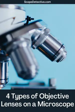
The four most common types of Microscope Objective Lenses
The below lenses are the four most common magnification levels for objective lenses. You can get lenses with other magnification levels also.
Scanning Objective Lens (4x)
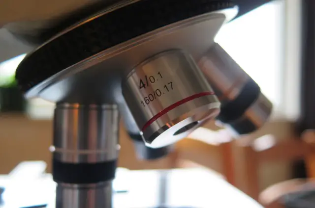
The scanning objective lens usually has 4x magnification and can be identified by a red strip band around the perimeter of the lens.
The scanning objective is designed for getting your bearings right before moving onto the low power lens. Its name, the ‘scanning’ lens, derives from the fact you are zoomed-out enough that you can scan around your specimen at this magnification level to prepare to move on to higher magnifications.
Using this objective, aim to:
- Achieve focus using the coarse focus knob.
- Center the specimen using your fingers or the mechanical stage.
A 4x magnification lens will usually achieve between 40x and 80x total magnification (with a 10x and 20x eyepiece respectively). This is the same sort of magnification that you’d get with a stereo microscope, and can provide close-up magnification of visible specimens such as feathers and leaves.
Low Power Lens (10x)
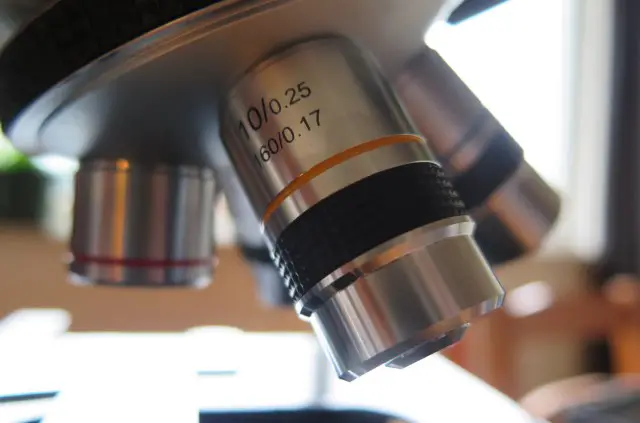
The low power objective lens usually has 10x or 20x magnification. You can identify it by a yellow strip around the lens housing.
This objective can be very useful for viewing prepared specimens on slides. It will achieve somewhere around 100x to 200x magnification for a 10x or 20x eyepiece respectively. This sort of magnification is great for viewing small specimens like:
- Fly legs
- Onion skin
- Brine shrimp
- Hair
- Skin
To use this lens, first start with the red striped scanning lens to achieve focus and center the specimen. Then rotate the objective turret clockwise to the yellow striped low power lens. Next, adjust the focus with the coarse focus knob to refine your focus and the mechanical stage to re-center your specimen.
High Power Lens (40x)
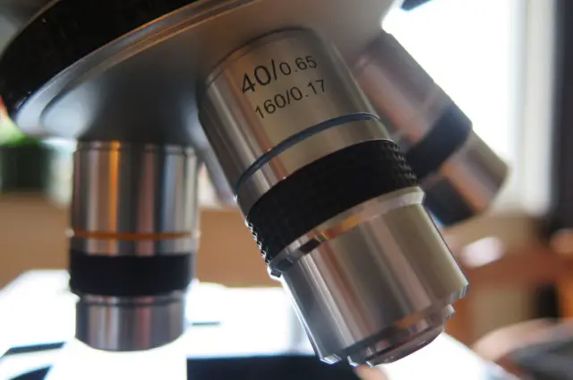
The high power lens is identifiable by a blue strip around the lens housing. Most compound light microscopes are sold with a 40x magnification high power lens, although this is not always the case. You may get a 32x or 60x magnification high power lens, for example.
The high power lens is used for looking at smaller specimens like bacteria and cells that are invisible to the naked eye. It’s commonly used for looking at:
- Mould
- Cells
- Germs
- Tardigrades
A 40x lens will achieve magnification of 400x when combined with a 10x eyepiece or 800x magnification with a 20x eyepiece. It is also very commonly used with 25x eyepieces to reach 100x magnification.
Oil Immersion Lens (100x)
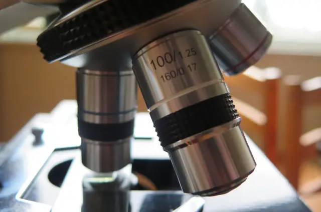
Not all beginner level microscopes will have an oil immersion lens, and they’re not really necessary for most hobby microscopy experiments.
You can identify a 100x lens because it will have a white or off-white cream colored stripe around the lens housing.
These lenses are called ‘oil immersion lenses’ because you usually need to use them with oil in order to view your specimen. At higher magnifications achieved with a 100x lens, there will often be too much distortion in the final image. To address distortion at high magnification levels, you can use oil immersion.
Oil immersion is achieved by placing a drop of oil above your specimen, then rotating the 100x lens over the oil so the gap between the specimen and the lens is covered by oil rather than air. The light waves moving through the oil will experience less distortion than if they move through air.
Total Magnification of a Compound Light Microscope
To identify the total magnification that you will achieve on a microscope, you need to multiply the magnification of the objective lens with the magnification of your eyepiece. Most microscopes come with 10x, 20x, or 25x eyepieces.
Here are the common total magnifications you can achieve with common light microscope settings:
|
|
10x Eyepiece |
20x Eyepiece |
25x Eyepiece |
|
4x Scanning Lens |
40x |
80x |
100x |
|
10x Low Power Lens |
100x |
200x |
250x |
|
40x High Power Lens |
400x |
800x |
1000x |
|
100x Oil Immersion Lens |
800x |
2000x |
2500x |
Specialty Objective Lenses for Advanced Microscopy Methods
There are some specialty objective lenses that you can also purchase for a microscope to conduct advanced microscopy methods. Four common specialty lenses are listed below.
1. Phase Contrast Objectives
Phase contrast is an advanced microscopy method designed to increase contrast for translucent specimens in order to make them more visible. A phase contrast objective has a dark ring around the lens to manipulate phase variations in light rays and convert them to amplitude variations. In simple terms, it manipulates light rays to deliver strong image contrast when looking through the eyepiece.
To conduct phase contrast methods, you need both a phase contrast objective and a specialized substage condenser.
2. Differential Interference Contrast (DIC) Objectives
Differential interference contrast (DIC) objectives are designed specifically to increase contrast of translucent specimens during brightfield microscopy. With a DIC objective, the need to use staining techniques is minimized as contrast can be achieved without staining.
A DIC objective is rarely found on a compound light microscope bought for hobby or school use.
3. Long Working Distance (LWD) Objectives
Long Working Distance objectives are designed to view specimens when the objective is a longer distance away from the specimen than usual. This is often required when a specimen is embedded in thick slides or below thick glass plates.
You can get other long working distance objectives such as LWD (Long Working Distance), ULWD (Ultra-Long Working Distance), and ELWD (Extra-Long Working Distance).
4. Reflected Darkfield Objectives
A reflected dark field objective is designed specifically for darkfield microscopy techniques. These techniques create a black background with high contrast to help to see specimens that are hard to see due to their translucency.
For a reflected darkfield objective, it is a lens structure also designed specifically for viewing specimens that are not placed within a covered slide.
You can tell you have a reflected darkfield objective because it will have demarcations such as Neo, BF/DF, or BD written on the objective. See here for more information on Reflected Darkfield Objectives.
Types of Objectives with Aberration Correction
1. Achromatic Lenses
Most microscope objectives are achromatic, to the extent that if there are no markings stating that they are not achromatic, you can assume that they are. In otherwise, this is the standard lens type. An achromatic objective normalizes red and blue light so they meet at the one focal point, while also correcting green light for spherical aberrations.
2. Apochromatic Lenses
These lenses are adjusted for blue, green, red and also deep blue. They are more expensive than chromatic lenses but also produce better high color images for the eye. They’re usually also spherically corrected for blue color aberrations.
3. Plan
Plan objective lenses are designed to correct for spherical aberrations to produce a crisp flat image. Plan corrections can be combined with other lens types, so you can have plan-achromatic, plan-apochromatic, etc. You can also have semi-pan objectives.
4. Infinity Corrected Lenses
Infinity corrected lenses allow for a theoretically infinite distance between the front and back of the objective. DIN standard objectives have a 160mm distance between the objective’s focal plane and back of the lens, and these lenses are often calibrated to perfectly align for that 160mm distance. By contrast, infinity correction projects a parallel light beam to the back of the objective. This is hard to explain, but easy to understand if you look at the diagram on this page.
Oil vs Dry Objectives
1. Dry Objectives
By default, compound light microscopes have dry objectives, meaning the space between the specimen and the objective is simply filed with air. Any objective with magnification under 100X you can assume is a dry objective unless otherwise marked.
2. Oil Immersion Objectives
100X objectives and above are often designed to allow for oil immersion. These are called ‘oil’ or ‘wet’ objectives. You can place oil in the space between the specimen and the objective so the light waves passing through that space pass through oil rather than air. This decreases distortion and leads to a higher quality image.
Oil immersion is often necessary at higher magnifications because of the physical limits of magnification. At super high magnifications, the light moving through air distorts too much to create a quality image. Oil immersion can minimize this issue.
Standards for Microscope Objective Lenses
1. DIN Standard
DIN is the international standard for objectives which regulates several elements of the objective, including the thread size. In practice, a microscope that accepts DIN standard objectives is incredibly versatile because you can buy replacement objectives without too much hassle. You just need to buy an objective that complies with DIN standards.
2. Color Bands
The color bands of objectives are more or less universal. They are:
- Red: 4x magnification
- Yellow: 10x magnification
- Blue: 40x magnification
- White: 100x magnification
There are other colors for less common objectives. See: objective lens color codes.
3. Numerical Aperture
You will find that the numerical aperture (NA) of the objective is usually demarcated on many objective lenses. Ensure you NA of the objective and that of the condenser are compatible for best quality image.
Conclusion
As you can see, there are many different types of objective lenses on microscopes. This list is by no means exhaustive, but does outline the common objective types that you will find on a regular compound light microscope in a school or home setting.
In reality, 90% of all beginner to intermediate experiments can be conducted with the four objectives you get out of the box – these are: the scanning objective, low power objective, high power objective and oil immersion objective.

Hi, I’m Chris and I run things around here! I share all my microscopy experiments, microscope information and tricks, how to guides, and microscope reviews in the articles on this site. Browse around to see what you like (I recommend the experiment ideas section) or connect with me on any of the social platforms listed below.